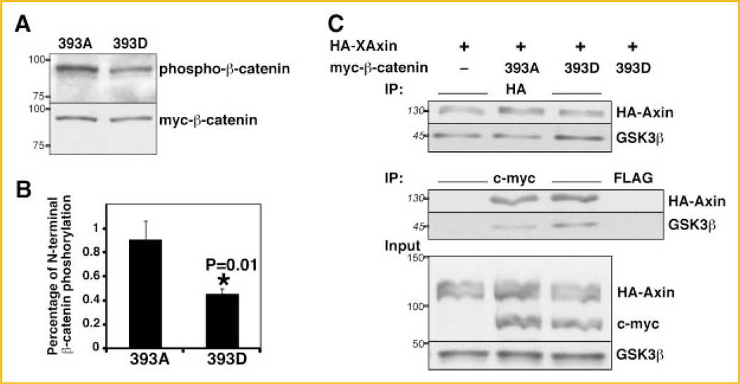
Fig. 6.
Reduced phosphorylation of 393D β-catenin by CKI/GSK3β. A: C57MG cells were lysed 24 h after transfection with 393D or 393A myc-β-catenin or pCS2 (not shown). Cell lysates were subjected to immunoblotting with the indicated antibodies. B: Histogram representing quantitation from three independent experiments showing the relative N-terminal phosphorylation of myc-β-catenin mutants relative to their expression. Asterisk (*) indicates P < 0.05. C: Higher affinity of endogenous GSK3β for Axin in HEK293T cells expressing 393D β-catenin. The experimental protocol was the same as in Figure 4C. Expression levels of exogenous and endogenous proteins are shown in the lower panel (input). Endogenous GSK3β levels are not affected by β-catenin transfection. Representative immunoblots from four independent experiments. Indicated on the left of the immunoblots are the apparent molecular weights of the proteins (numbers in italics) or the position and weight of the molecular markers utilized (numbers in roman).

