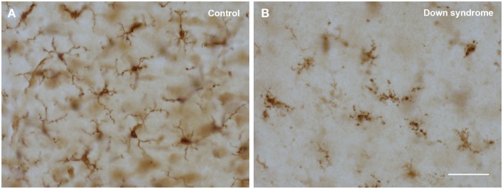
Figure 3.
Comparison of normal (ramified) and degenerating (dystrophic) microglia using Iba1 immunostaining in human cerebral cortex. (A) 22-year-old male non-demented subject reveals cells with normal morphology; (B) 48-year-old female subject with Down syndrome shows cells displaying obvious cytoplasmic fragmentation. Scale bar: 50 μm.

