Figure 3.
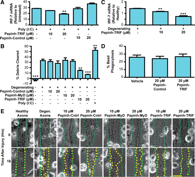
Axonal debris is cleared by microglia in a TRIF-dependent manner. A, Rat microglia were preincubated with peptide inhibitors, followed by treatment with poly(I:C). Real-time PCR of IRF7 shows that TRIF inhibition by Pepinh-TRIF decreases IRF7 expression at 16 h. B, Quantification of axon debris clearance at 16 h demonstrates that TRIF inhibition markedly reduces axon debris clearance in a dose-dependent manner. One-way Tukey's ANOVA analysis, comparisons with degenerating condition; F value, 18.31. C, Real-time PCR of microglia cocultured with degenerating axons demonstrates that TRIF inhibition results in decreased IRF7 expression. D, TRIF inhibition does not alter bead phagocytosis. A total of 1 × 10⁶ FluoSpheres was added to microglia in 96-well plates. The percentage of microglia that took up more than one bead was determined. Error bars indicate SEM. **p < 0.01; ***p < 0.001. E, Representative images of debris clearance. The dotted green traces outline axon bundle areas at time 0, and the dotted yellow traces outline the axon bundles after 16 h. Scale bar, 25 μm.

