
Figure 6.
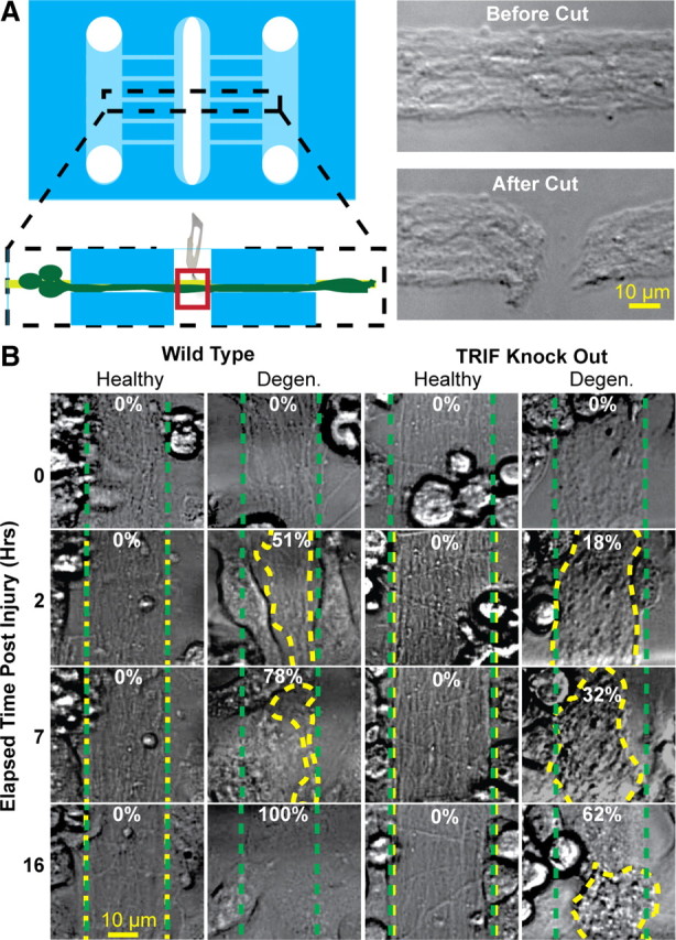
Microglia clear axonal debris following axotomy. A, The microfluidic platform was modified by creation of a third compartment. Before addition of wt or TRIF KO mouse microglia, a scalpel was used to sever a 10–20 μm segment of the bundle, thereby inducing axon degeneration to distal axon segments. B, Mouse microglia were then cocultured with either healthy or cut axon bundles to assess the extent of axon debris clearance. The dotted green traces outline axon bundle areas at time 0, and the dotted yellow traces outline the axon bundles after indicated times. The numbers refer to the percentage clearance of degenerating axons.