
Figure 7.
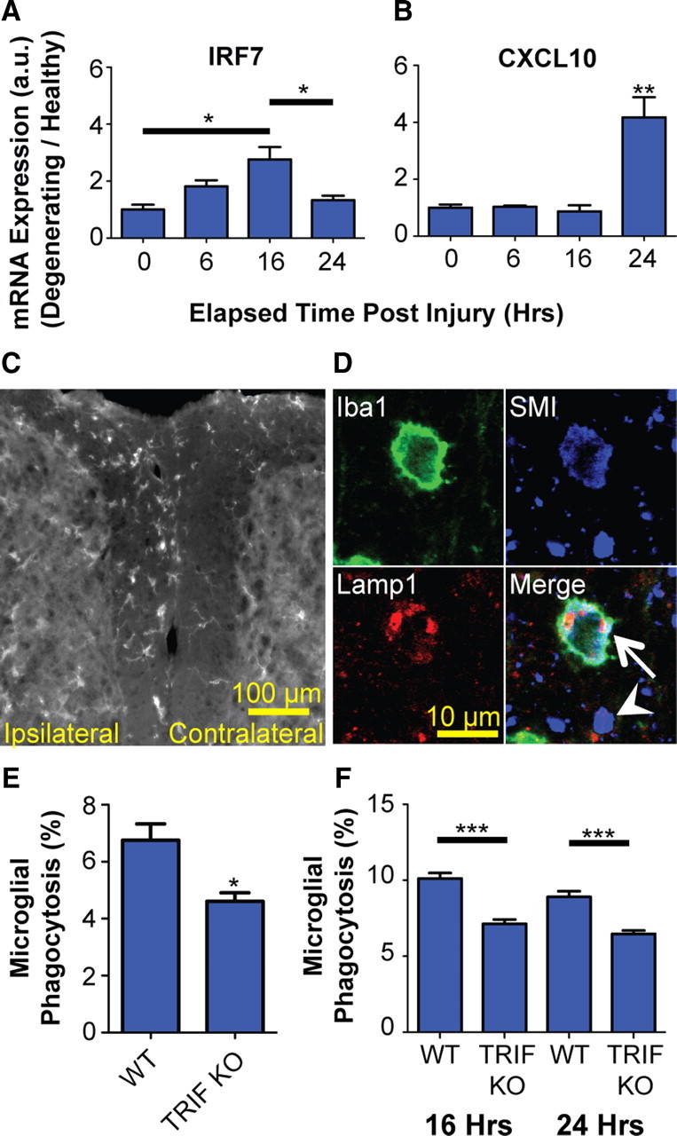
TRIF KO impairs microglial phagocytosis in vivo. Real-time PCR of ex vivo isolated microglia following dorsal root axotomy demonstrates induction of IRF7 (Tukey's ANOVA; F value, 7.95) (A) and CXCL10 (Tukey's ANOVA; **p < 0.01 compared with no axotomy control, labeled “0”; F value, 18.16) (B). C, Dorsal root axotomy results in microglial accumulation in ipsilateral dorsal column within 2 d of injury. Scale bar, 100 μm. D, Triple-label immunohistochemistry and confocal microscopy of ipsilateral side show neurofilament-positive axons (SMI) colocalized with endosomes (Lamp1) within microglia (Iba1). Arrow, Microglia phagocytosing axon; arrowhead, extracellular (nonphagocytosed) axonal material. Scale bar, 10 μm. E, Quantification of axon phagocytosis by microglia (Iba1+ SMI+ cells) in ipsilateral dorsal columns at 2 d following axotomy. *p < 0.05, Student's t test. F, In vitro quantification of axon phagocytosis by wt or TRIF KO microglia cocultured with TdTomato-labeled axons. ***p < 0.001, Tukey's ANOVA analysis; F value, 28.23. Error bars indicate SEM.