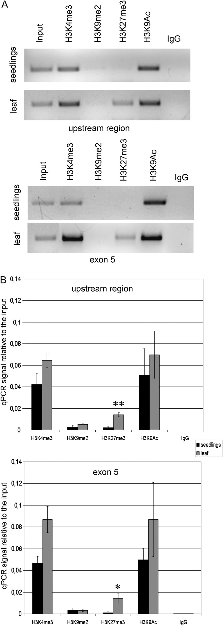
Fig. 4.
Analysis of histone modifications in the AtTERT upstream region and in exon 5 by ChIP. DNAs from immunoprecipitated fractions of chromatin were purified and a 336 bp region upstream of the ATG signal and a 476 bp region of the fifth exon were amplified using classical (A) or quantitative (B) PCR (qPCR). (A) A representative example of PCR amplification of the AtTERT upstream region and of exon 5 in immunoprecipitated fractions. Signals of euchromatin-specific marks (H3K4me2, H3K9Ac) were strong in both tissues analysed; signals for the modification typical for constitutive heterochromatin (H3K9me2) were below the detection limit. Note the distinct H3K27me3 band in the leaf samples. (B) Two biological replicates of wild-type seedlings and mature leaves were immunoprecipitated and subjected to quantitative PCR. Signal from the immunoprecipitated fractions was expressed relative to that from the total input chromatin. The amount of the H3K27me3 mark increased in the telomerase-negative tissue (leaf) in both regions analysed (P < 0.01 in the AtTERT upstream region; P < 0.05 in the exon 5).

