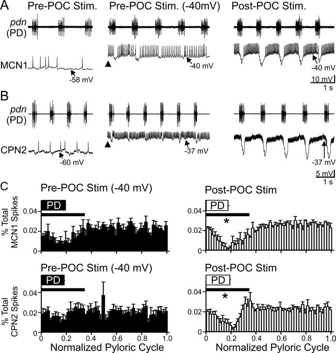
Figure 3.
CPG feedback to projection neurons is strengthened by POC stimulation. A, B, Left, At their resting potentials before POC stimulation, AB inhibition is weak in MCN1 and CPN2. Middle, Before POC stimulation, MCN1 and CPN2 were depolarized via intracellular current injection to approximately −40 mV (arrowheads). Under this condition, AB inhibition elicits small-amplitude hyperpolarizations in MCN1 and CPN2 that modestly regulate their firing rate. Right, POC stimulation depolarizes MCN1 and CPN2 to approximately −40 mV, during which time AB inhibition elicits larger amplitude hyperpolarizations and longer duration pauses in their firing, despite their firing rate being increased (MCN1) or comparable (CPN2) relative to their activity during the current injections before POC stimulation (see Results). MCN1 and CPN2 recordings are from different preparations. C, The percentage of MCN1 (top) and CPN2 (bottom) action potentials per bin (2% of pyloric cycle per bin) and the phase of AB/PD activity (PD box) are plotted against the normalized pyloric cycle (see Materials and Methods) before (left) and after (right) POC stimulation. The pre-POC stimulation data were obtained while MCN1/CPN2 was depolarized by continual current injection to approximately −40 mV. After POC stimulation, there is a larger decrease in the percentage of MCN1/CPN2 spikes during the initial 35% of the normalized pyloric cycle (bar), relative to pre-POC stimulation. Means ± SEM per bin are plotted. MCN1: n = 4; CPN2: n = 4. *p < 0.05.

