
Figure 2.
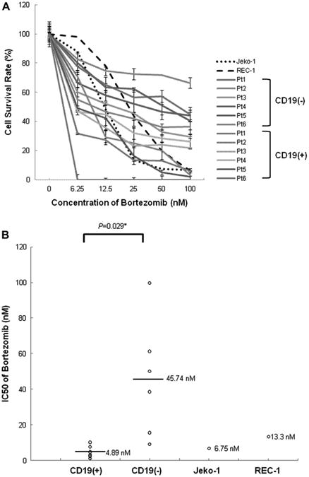
CD45+CD19− MCL-ICs isolated from patient samples are bortezomib-resistant. (A) The bortezomib sensitivities of CD45+CD19− MCL-ICs and CD45+CD19+MCL cells isolated from different six MCL patients were compared with those of REC-1 and Jeko-1 cells. Cells (1.5–2.5 × 10⁵ cells per well) were cultured in 24-well plates for 16 hours after the addition of bortezomib. Cell viability was determined by CellTiter-Blue fluorometric assay (Promega) and was indicated as a ratio compared to cell viability without treatment. Drugs were serially diluted as indicated from maximum drug doses of 100 nM. Results show the mean ± standard deviation of triplicate. CD45+CD19− MCL-ICs showed more survival rates on all tested drug concentrations compared to CD45+CD19+ MCL cells. CD19(−), CD45+CD19− cells; CD19(+), CD45+CD19+ cells. (B) The mean IC50 value of bortezomib for CD45+CD19− MCL-ICs was much higher than those of the CD45+CD19+ MCL cells, Jeko-1, or even REC-1, which was reported as a bortezomib-resistant cell line. CD19(−) = CD45+CD19− cells; CD19(+) = CD45+CD19+ cells. Bars represent averages; *p < 0.05 by unpaired t-test.