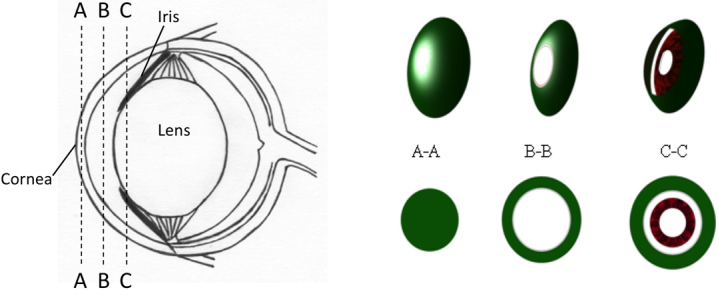
Figure 2.

Diagram showing the different regions of the mouse eye that were measured with the multiphoton microscope, results shown in Figure 3. A-A represents a solid cut of the cornea from the top surface into the stroma. B-B is a cross-section of the anterior chamber filled with aqueous humor. Since the aqueous humor does not emit any signal, the sectioned image appears in the shape of a ring. C-C is a section deep inside the anterior chamber that includes the front of the iris.
