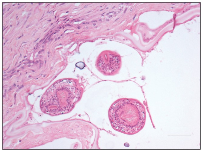
Figure 3.
Photomicrograph of a section of cystic tissue from the mass shown in Figure 2. Note the 3 intraluminal protoscolices surrounded by a thin membrane. The cyst is lined by a hyaline membrane (“laminated layer”). Hematoxylin and eosin, Bar = 50 μm.

