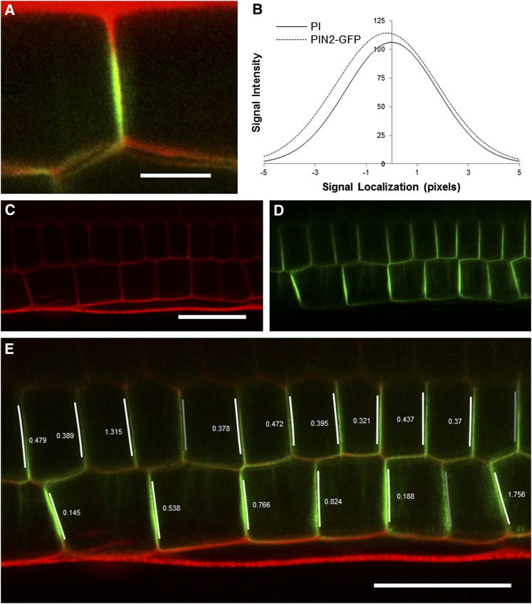
Figure 4.
Output of the Super-Resolution Plug-in.
(A) A zoomed section of a confocal image showing the PIN2-GFP protein residing on one side of an epidermal anticlinal cell wall. Red channel: cell walls stained with propidium iodide. Green channel: PIN2-GFP.
(B) Example plot of two normal distributions fitted to the wall and PIN2-GFP channels of (A). The offset (0.195 pixels; 0.04 μm) of the means of these two distributions reveals which side of the wall the protein resides.
(C) and (D) Input image of epidermal and cortical cells files of a root expressing PIN2-GFP.
(E) Output of protein localization plug-in with the direction and size of the offset (in pixels) of the PIN2-GFP channel overlaid in epidermal and cortical cell files. Overlaid arrows indicate rootward and shootward polarities. In three locations, no value is shown where this offset is below the detection limit set at 0.1 pixels, and so would not be used for further analysis. PIN2-GFP polarity is correctly detected as shootward in all epidermal cells and rootward in the majority of cortical cells. See also Supplemental Movie 3 online.
Bar in (A) = 5 μm; bar in (C) = 30 μm; bar in (E) = 30 μm.

