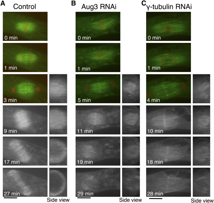
Figure 6.
Phragmoplast MT Formation Is Severely Suppressed by Augmin or γ-Tubulin RNAi.
Time-lapse imaging of a caulonemal cell expressing GFP-tubulin (green) and histone-RFP (red) by spinning-disk confocal microscopy. Images were acquired every 1 min, each with 13 z-sections (separated by 1 µm), and are displayed after maximum projection. A hypomorphic line (#24) was used for γ-tubulin in this experiment, which entered anaphase more frequently (five of eight anaphase cells showed this phenotype). Note that a cell was not entirely covered by 13 sections, so that an incomplete ring is seen after projection of the control cell. Also see Supplemental Movies 5 and 6 online. Bars = 10 µm.

