Figure 1.
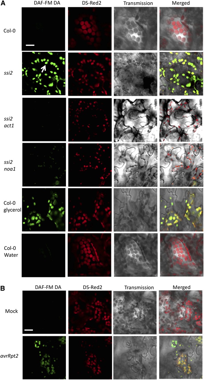
The ssi2 Plants Accumulate High Levels of Chloroplastic NO.
(A) Confocal micrograph of DAF-FM DA–stained leaves showing subcellular location of NO in wild-type (Col-0) plants, ssi2, ssi2 act1, and ssi2 noa1 mutants, and glycerol-treated Col-0 wild-type plants. Chloroplast autofluorescence (red) was visualized using Ds-Red2 channel. Arrow indicates chloroplast. At least 10 independent leaves were analyzed in four experiments with similar results. Bar = 10 μm.
(B) Confocal micrograph showing pathogen-induced NO accumulation in Col-0 plants at 12 h after inoculation. Plants were inoculated with MgCl2 (mock) or avrRpt2 P. syringae. At least 10 independent leaves were analyzed in four experiments with similar results. Bar = 10 μm.

