
Figure 2.
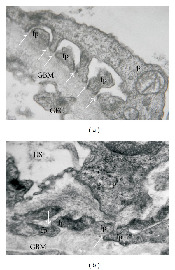
Glomerular filtration barrier in normal and pathological conditions. (a) A cross-section of a normal human glomerular capillary. Arrows indicate the foot processes with interconnecting ultrathin slit diaphragms. (b) Significant changes in the glomerular filtration barrier in a patient with heavy proteinuria due to a primary glomerulopathy. Note the foot process effacement with alteration in slit diaphragm (arrows), which results in a disarrangement of the functional filter structure. Abbreviations: fp: foot process; GBM: glomerular basement membrane; P: podocyte; GEC: glomerular endotelial cell; US: urinary space. (Transmission electron microscopy. Uranyl acetate-lead citrate original magnification ×40,000.)