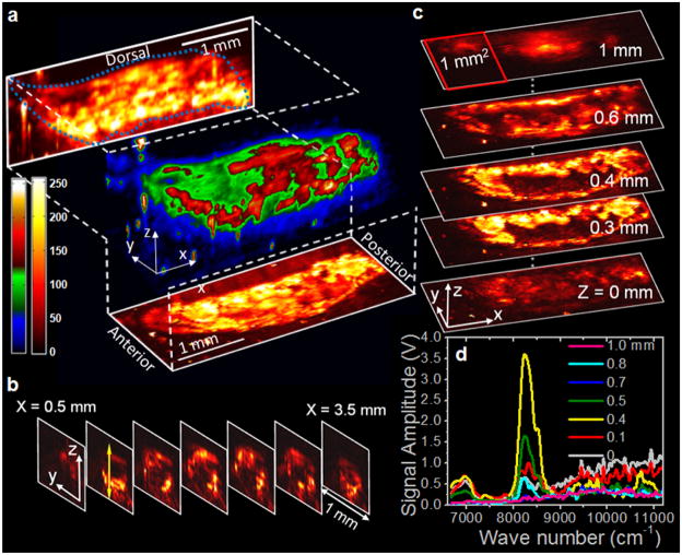
FIG. 3.
(color online). In vivo 3D VPA imaging of fat bodies in a 3rd-instar larva of Drosophila melanogaster. (a) 3D reconstruction and maximum amplitude projections in XZ and XY planes, (b) transverse images, and (c) longitudinal (planar) images of the lipid storages in a Drosophila larva. (d) Depth-resolved VPA spectra at a location indicated by the light gray (yellow) arrow in (b). 3D animation is available as a supplementary video; see [17].

