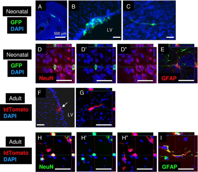
Figure 7.
NSCs from the SVZ are multipotent and neurogenic in vivo after transplantation. A, GFP-positive PD4-NSCs (green) 4 h after transplantation into PD0 mouse brains. B, GFP-positive cells (green) were observed in the lateral ventricle (LV) 1 month after transplantation. C, GFP-positive cells (green) with neuronal morphology were observed in the olfactory bulb 1 month after transplantation. D, E, Confocal imaging demonstrated that GFP-positive PD4-NSCs (green) differentiated into NeuN-positive neurons (D, red) and GFAP-positive astrocytes (E, red) in the olfactory bulb. D′, D″, FITC and TRITC channels, respectively. F, tdTomato-positive adult NSCs (red) derived from the SVZ were observed in the LV 1 month after transplantation. G, tdTomato-positive cells (red) with the morphology of migrating neuroblasts were observed in the RMS 1 month after transplantation. H, I, tdTomato-positive adult NSCs (red) differentiated into NeuN-positive neurons (H, green) and GFAP-positive astrocytes (I, green) in the olfactory bulb 1 month after transplantation. H′, H″, FITC and TRITC channels, respectively. Cell nuclei are shown by DAPI staining (blue). Scale bars: 500 μm where indicated; otherwise 50 μm.

