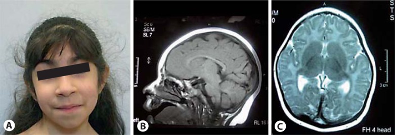
Fig. 1.
Photograph of patient 1 at the age of 5 years and 2 months (A). She shows only mild facial dsymorphism with upslanting palpebral fissures and broad tip of the nose. MRI scan of the brain at the age of 6 months shows small frontal lobes and mild ventriculomegaly, while the sizes of brain stem and cerebellum were normal (B, C).

