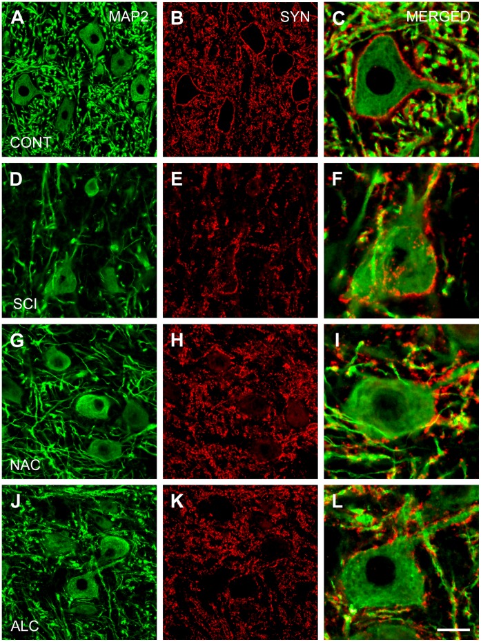
Figure 3. Expression of microtubule-associated protein-2 and synaptophysin.
Horizontal sections through the ventral horn of L4–L5 segments showing immunostaining for microtubular-associated protein-2 (MAP2; dendritic branches, left column) and synaptophysin (SYN; synaptic boutons, middle column) of a control animal (A–C; CONT), at 4 weeks after spinal cord injury (D–F; SCI) and following treatment with N-acetyl cysteine (G–I; NAC) or acetyl-L-carnitine (J–L; ALC). Note that synaptic boutons around motoneuron cell bodies are not recovered after NAC or ALC treatment (right column). Scale bar, 50 µm (left and middle columns) and 20 µm (right column).

