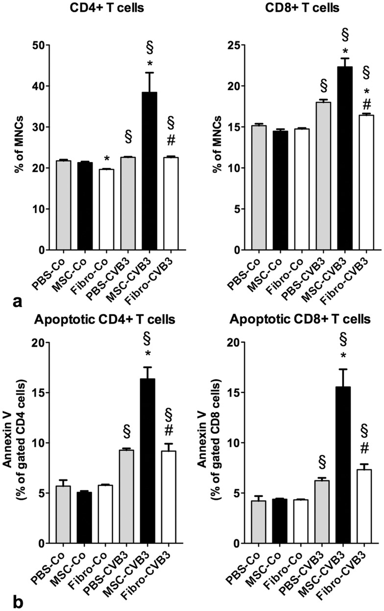
Figure 2. Flow cytometric analysis of splenocytes 7 days after infection.
Viral infection led to a significant increase of CD4+ and CD8+ T cells in the spleen, but also induced apoptosis in both cell types. Administration of MSCs led to a significant increase of CD4+ and CD8+ T cells in the spleen in the infected animals compared to the non-treated mice. However, MSCs also significantly increased the apoptosis rate of CD4+ and CD8+ T cells in the infected mice. *p<0.05 vs PBS-CVB3, § p<0.05 vs respective control-group, # p<0.05 vs. MSC-CVB3.

