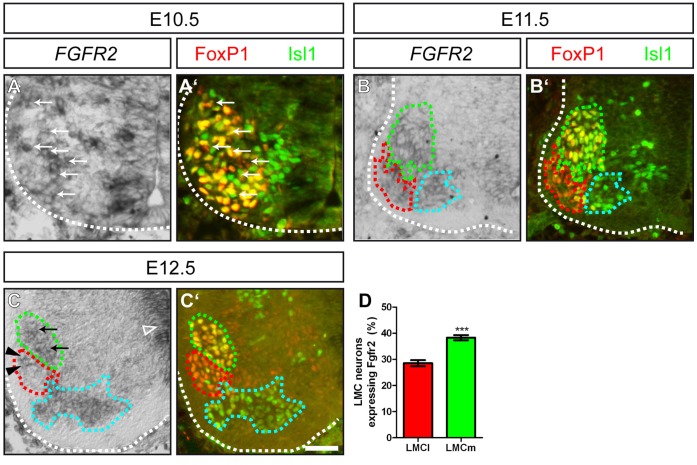
Figure 1. FGFR2 is expressed in motor neurons of the LMC during forelimb innervation.
(A, A’) At E10.5, spinal motor neurons in the ventral horn of the brachial spinal cord that will form the medial and lateral aspect of the LMC are identified by FoxP1 and/or Isl1 immunohistochemistry. A subset of these motor neurons shows expression of FGFR2 (arrows). (B, B’) At E11.5, motor neurons have segregated into two distinct sub-columns of the LMC; namely the LMCm (FoxP1+/Isl1+, green dashed line) and the LMCl (FoxP1+/Isl1−, red dashed line). FGFR2 mRNA is found in the LMC and MMC (FoxP1−/Isl1+, cyan dashed line). (C, C’) In situ hybridization against FGFR2 shows a higher number of motor neurons that express the FGF receptor in the LMCm (FoxP1+/Isl1+, green dashed line, arrows) when compared to dorsally projecting motor neurons of the LMCl (FoxP1+/Isl1−, red dashed line). (D) Quantification of FGFR2 mRNA expression in motor neurons of the LMCm and LMCl showed a significantly higher number of ventrally projecting motor neurons that expressed the FGF receptor. Scale bar in (C’) equals 25 µm for (A), 40 µm for (B) and 50 µm for (C).

