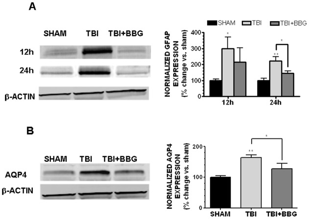
Figure 7. BBG attenuates glial activation.
(A) Representative Western blot (left panel) of cortical GFAP expression taken at 12h or 24h after sham injury, TBI, or TBI +50 mg/kg BBG. (B) Representative Western blot (left panel) of AQP4 in the cerebral cortex of mice at 12h following sham injury, TBI, or TBI +50 mg/kg BBG. Densitometric analysis of Western blots (right panels) is presented as either GFAP or AQP4 expression following normalization to β-actin, which was used to control for equal protein loading. Data (mean ± SEM) are representative of six mice/group from three independent experiments (n = 3/group in each experiment) and are expressed as % change vs. sham. Data were analyzed by One-Way ANOVA followed by Dunnett's post-hoc test (* p<0.05, ** p<0.01 vs. sham operated mice).

