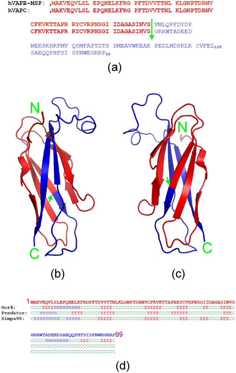
Figure 1. Comparison of VAPC and VAPB-MSP.
(a) Sequence alignment of the human 99-residue VAPC and 125-residue VAPB-MSP domain. The first 70 residues (in red) are completely identical, while the rest (in blue) are different in the two proteins. The green arrow is used to separate the first 70 residues from the rest. (b, c) Crystal structure of the 125-residue VAPB-MSP domain that we previously determined (46), in which the 70 identical residues are in red, while the different ones are in blue. (d) Secondary-structure prediction of VAPC by computational programs GOR4, Predator, and SIMPA96. Red E, β-strand; blue H, helix; green C, random coil.

