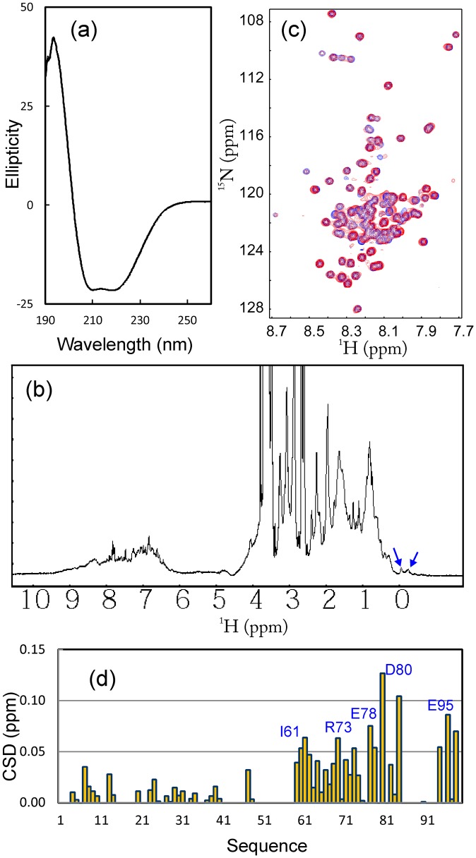
Figure 3. Binding between VAPC and NS5B.
(a) Far-UV CD spectrum of HCV NS5B. (b) One-dimensional 1H NMR spectrum of HCV NS5B. Blue arrows are used to indicate very up-field NMR resonance peaks characteristic of a well-folded protein with tight tertiary packing. (c) Superimposition of 1H-15N NMR HSQC spectra of VAPC in the absence of (blue) and in the presence of unlabeled NS5B (red) at a molar ratio of 1∶1.5 (VAPC:NS5B). (d) Residue-specific changes of integrated 1H and 15N chemical shifts of VAPC in the presence of unlabeled NS5B at a molar ratio of 1∶1.5 (VAPC:NS5B).

