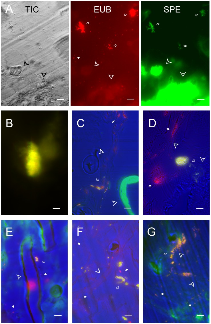
Figure 3. Bacterial localization in the gut of S. littoralis larvae with Fluorescent In Situ Hybridization.
Scale bar equals 10 µm. A, Detection of Clostridium sp. In the midgut. The three images shown are TIC image, fluorescent image of universal probe (EUB, red) and of specific probe (SPE, green). B to G are merged images of TIC, EUB and SPE. The bacteria detected only with universal probe are red, and the bacterial with both probes are green. B, a large aggregate of Clostridium sp. deep in the gut lumen. C, Detection of E. mundtii. D, Detection of E. casseliflavus. E, P. acnes in the midgut. F, E. coli detected in the midgut; G, K. pneumonia detected in the midgut. Bacteria detected only by universal probe are highlighted with white arrows; Bacteria stained by sequence-specific probes are pointed by open arrows. Insect tissue is indicated by arrow heads.

