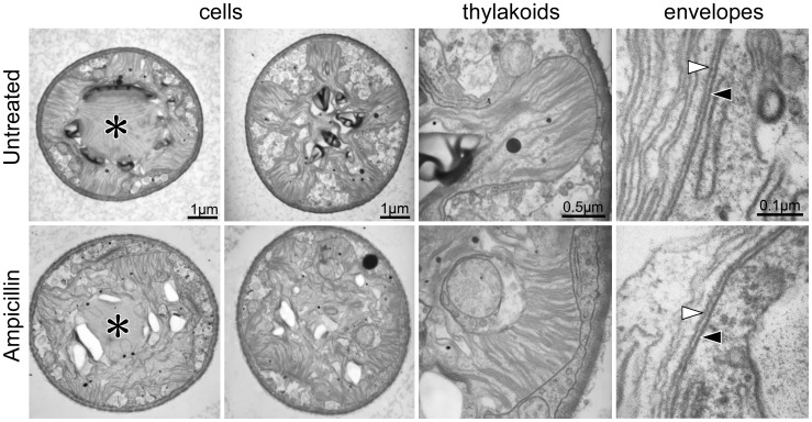
Figure 3. Electron micrographs of ampicillin-treated Closterium cells and of untreated controls.
The magnification in the lower photos is the same as that in the upper photos. Pyrenoids surrounded by starch are indicated by asterisks. Black and white triangles indicate the outer and inner envelopes of chloroplasts, respectively.

