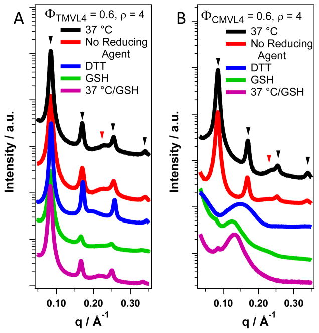
Figure 5.
SAXS data for complexes prepared from TMVL4 and CMVL4 (ρ = 4, ΦDOPC = 0.4) incubated at different temperatures and with reducing agents. Black arrowheads mark peaks resulting from the lamellar ordering; red arrowheads mark the DNA correlation peak. (A) The scattering patterns of TMVL4-based complexes do not change upon addition of either reducing agent (DTT and GSH) or change in temperature, i.e., the complexes remain lamellar. Incubation at 37 °C was for 8 hours. The sample containing GSH was incubated an additional 5 months at 4 °C (magenta curve). (B) The scattering patterns of CMVL4-based complexes are strongly affected by the addition of reducing agents but not by incubation at physiological temperature without reducing agent. The peaks characteristic for the lamellar structure disappear completely (DTT) or almost completely (GSH) and a new broad peak at q = 0.15 Å−1 (DTT) or q ≈ 0.13 Å−1 (GSH) appears. The sample incubated with GSH at 4 °C for one month also shows a faint broad peak at q = 0.22 Å−1 which is absent in the sample incubated at 37 °C. Incubation at 37 °C was for 8 hours in all cases.

