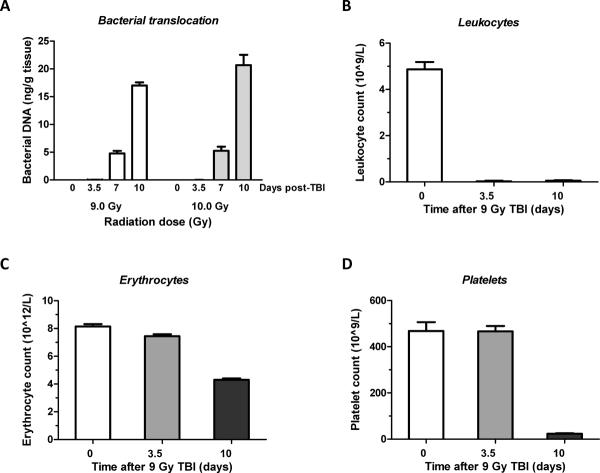
Figure 3. Bacterial translocation to the liver and peripheral blood cell counts after exposure to TBI.
-
A)Bacterial translocation was determined as bacterial load in liver tissue at baseline and 3.5, 7, and 10 days after exposure to 9 Gy or 10 Gy TBI.
- Significant amounts of bacterial DNA observed on day 7 (p=0.0006) and on day 10 (p=0.000005). Translocation was significantly greater after 10 Gy than after 9 Gy (p=0.01).
- Mean ± SEM, 4 mice per group (2 mice died before day 10 in the 10 Gy exposure group).
-
B–D)Changes in the level of blood cells were assesses at day 0, 3.5 and 10 after exposure to 9.0 Gy TBI.
- Leukocytes exhibited a significant decrease at 3.5 days and the erythrocyte count was borderline significantly decreased. At 10 days after TBI, all peripheral blood count parameters were highly significantly reduced.
- Mean ± SEM, 4–6 mice per group.

