
Figure 5.
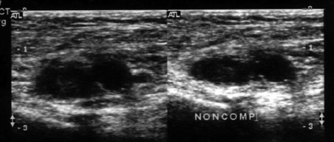
Transverse gray scale images of the common femoral vessels with acute deep venous thrombosis. The image on the left is without compression and the image on the right demonstrates the non-compressible vein and adjacent artery
Official websites use .gov
A
.gov website belongs to an official
government organization in the United States.
Secure .gov websites use HTTPS
A lock (
) or https:// means you've safely
connected to the .gov website. Share sensitive
information only on official, secure websites.
Transverse gray scale images of the common femoral vessels with acute deep venous thrombosis. The image on the left is without compression and the image on the right demonstrates the non-compressible vein and adjacent artery