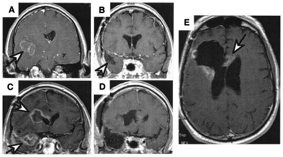
Figure 2.
MRI scans of a patient with a right temporal GBM illustrating the spread of the disease. (A) Presurgical scan, GBM (arrow) is surrounded with edema. (B) Scan after surgery and radiation therapy showing “gross total resection” and clear resection cavity, and (C) six months later, showing recurrence not only at the resection margin (arrow) but a second focus of GBM across the Sylvian fissure in the frontal lobe (arrow). (D) Postresection scans of both recurrent tumors. (E) Scan 3 months later, showing the tumor recurring at the resection margin and crossing the corpus callosum to the other hemisphere (arrow).

