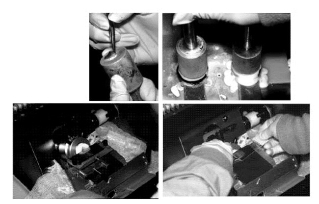
Figure 12. A. The first specimen is placed deep side down on a heat extractor and the epidermis is “teased” radially.B. The microtome chuck is prepared with OCT and the heat extractor is placed on to the chuck.C. The chuck is placed into the microtome and sectioned in the cryostat.D. The serial sections are placed sequentially on microscope slides.
