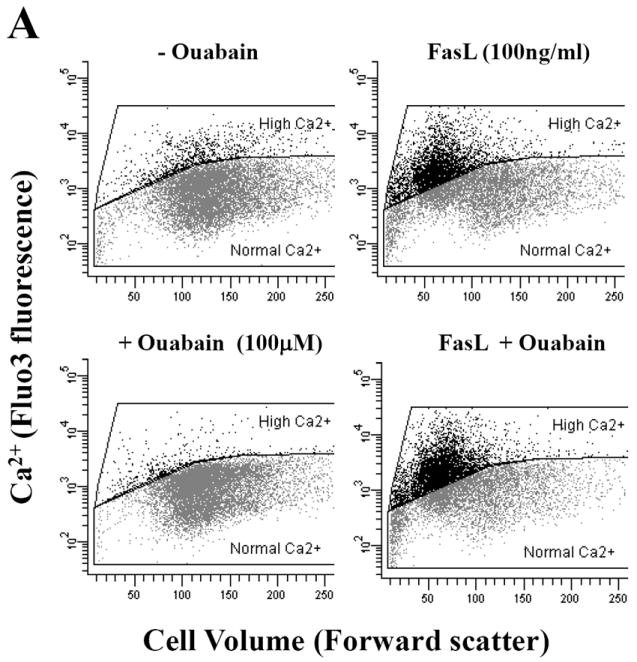
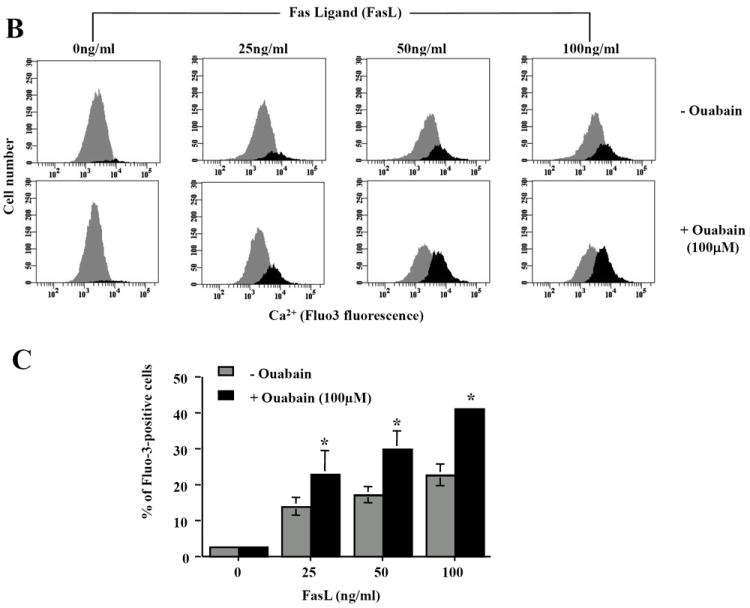
Fig. 4. Effect of ouabain on cell Ca2+ homeostasis in FasL-induced apoptosis.


Apoptosis was induced in Jurkat cells by addition FasL for 4h. Cells were loaded with Fluo-3 AM 30min prior to flow cytometry analysis. Intracellular Ca2+ content was analyzed in either Fluo-3 fluorescence versus forward scatter dot plots (A) or Fluo-3 fluorescence histograms (B). Shown are either a single representative experiment from three independent experiments, (A and B), or averaged values ± SEM from three independent experiments (C). Asterisks (*) indicate statistical significance (P<0.05) between the two experimental conditions at each concentration of FasL.
