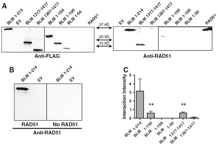
Fig. 2.

Far western Immunoblotting of BLM termini. a Left panel Conventional western analysis of the BLM fragments using anti-FLAG M2 antibody. Right panel Far western analysis of BLM fragments interaction with RAD51 using anti-RAD51 antibody. b Left panel Far western of BLM fragments incubated with RAD51 cell lysate. Right panel Far western of identical blot incubated with empty vector control cell lysate. c Quantification of data represented in (a). RAD51 far western interaction bands were normalized for BLM fragment loading controls. Graph represents the mean of three independent trials with standard deviations indicated. One-way ANOVA analysis was completed for all fragments with Dunnett’s multiple comparison test. Statistical significance of p < 0.01 is indicated with double asterisks
