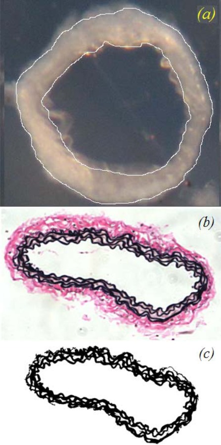
Fig. 5.

Images used for dimension and component measurements: (a) representative cut arterial ring with the inner and outer diameter outlined, (b) VVG stained histology section which clearly shows the boundaries of the media and adventitia in the artery wall, and (c) VVG stained histology image thresholded to highlight the elastic lamellae for area fraction measurements
