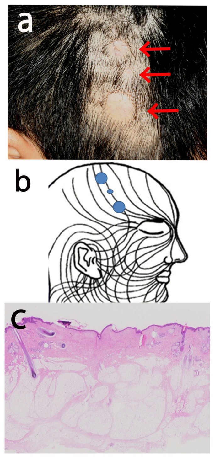
Figure 1.

Clinical and histological features. a: There are 3 atrophic scar-like alopecic macules linearly arranged on the right temporal area (arrows). b: Distribution of skin lesions in the present case transposed onto Blaschko’s lines as proposed by Happle et al (2). c: Histological section showing atrophic scar formation, namely, thin epidermis with a reduced number of rete ridges, proliferation of collagen fibers in the dermis, and absence of hair follicles orphaned arrector pili muscles and sweat glands. Original magnification, 10x.
