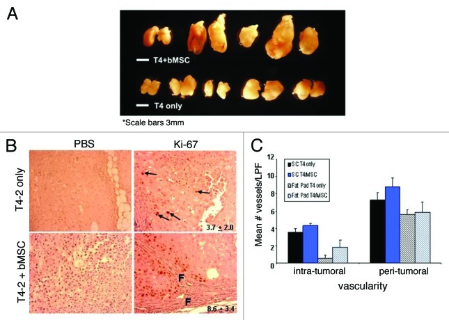
Figure 5. Breast MSCs promote the growth but not vascularity of HMT-3522 T4-2 cells. (A) 8-week HMT-3522 T4-2 cells (T4-2) ± bMSC3 fat pad xenografts (scale bar: 3mm). (B) Determination of the percent of Ki-67 positive cells per microscopic field revealed a statistically significant difference (p ≤ 0.011) between the pure and mixed xenografts. Representative immunohistochemical staining reveal that pure T4-2 xenografts contained only rare Ki-67 positive cells (arrows in top panels), whereas T4-2/bMSC xenografts often contained focal areas of active proliferation in close association with fibrosis (f = fibrosis). Primary antibody was omitted as a negative control for all specimens (left panels). Values indicated in the right panels represent mean percent Ki-67 positivity and were calculated based on counting the number of immunolabeled cells and then dividing by the total number of cells in 3 random fields per xenograft. (C) Intra- and peri-tumoral vascularity was not significantly different in T4-2 vs. T4-2/bMSC mixed xenografts whether injected orthotopically (patterned bars) or subcutaneously within Matrigel® (solid bars). Vascularity was determined on H&E stained tissue sections as described in Materials and Methods. LPF: low power field.

An official website of the United States government
Here's how you know
Official websites use .gov
A
.gov website belongs to an official
government organization in the United States.
Secure .gov websites use HTTPS
A lock (
) or https:// means you've safely
connected to the .gov website. Share sensitive
information only on official, secure websites.
