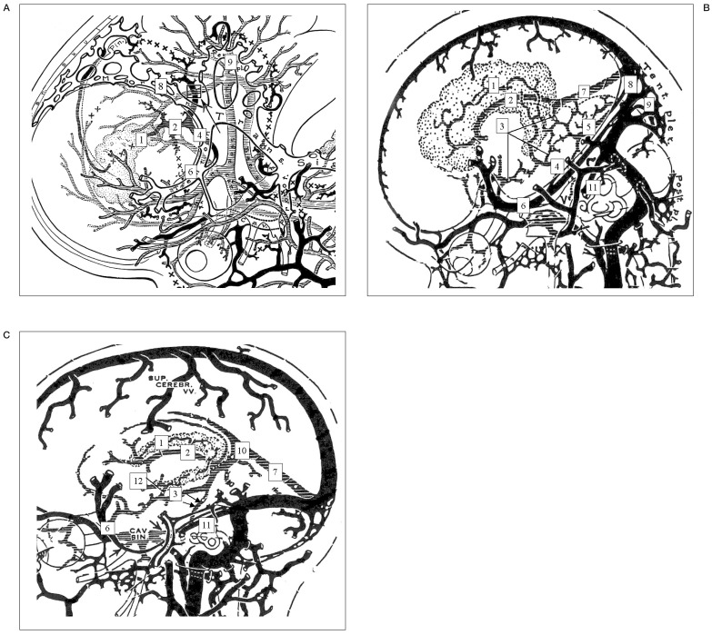
Figure 3.
Embryologic draining (anastomotic) patterns between the BVR and remnant embryonic tentorial sinus according to each stage of development by Padget DH7. A) Stage 6, 22-24 mm stage. Primarily the ventral diencephalic veins, supplemented by the primitive internal cerebral vein, drain the conspicuous choroid plexus of the lateral ventricle. B) Stage 7a, 60-80 mm stage. The internal cerebral vein is still primitive and constitutes the drainage of the large superior choroid vein into the straight sinus. The basal cerebral vein is formed by longitudinal anastomoses between several primitive pial veins, the deep middle cerebral, the ventral or dorsal diencephalic veins, the mesencephalic vein, and a tributary of the primitive straight sinus. C) Infant or adult stage. A lateral mesencephalic vein (LMV; arrows) anastomosing the basal veins with the ventral metencephalic veins sometimes become the only outlet of the basal veins.
1,superior choroidal vein;2,primitive internal cerebral vein; 3, primitive BVR; 4, ventral diencephalic vein; 5, mesencephalic vein, 6, embryonic tentorial sinus; 7, straight sinus; 8, marginal sinus (primitive transverse sinus); 9, tentorial plexus; 10, vein of Galen; 11, superior petrosal sinus (great anterior cerebellar vein); 12, inferior ventricular vein.

