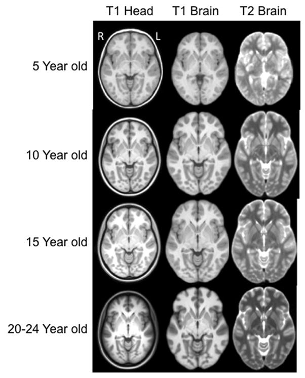
Figure 4.

Head and brain templates with T1W and T2W weighted images, for 5, 10, 15 and 20–24 years of age. The displayed data is based on final averaged output as outlined in Figure 1. (axial slices at AC-PC level). Each figure is show approximately as the same size and not scaled according to the actual age head size. Right of head is on left of figure.
