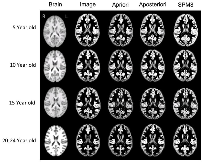
Figure 5.

Brain template (left column) and associated gray matter tissue probability maps (in the four right columns) based on segmentation methods used for 5, 10, 15 and 20–24 year old templates (axial slices at AC-PC level). Figures are not to scale with actual head size; right of head is on left of figure.
