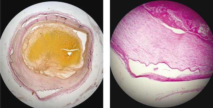
Figure 5.
EVG stain and H & E stain. A low power and high power view of the distal middle cerebral artery showing organizing thrombus within the lumen, an irregularly thickened intima and disruption of the internal elastic lamina. Poor cohesion between internal elastic lamina and adventitia creates artefactual splits.

