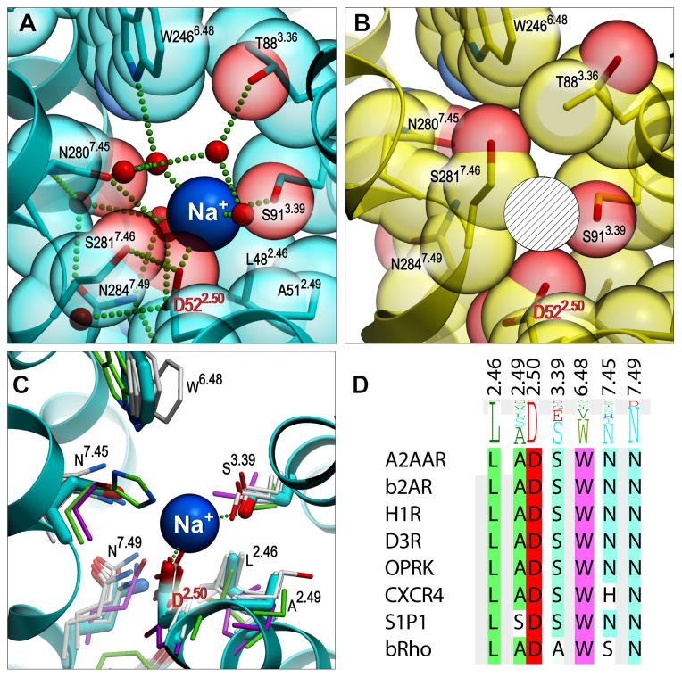
Fig. 2. Structural details of the Na+ allosteric site in the inactive and active-like state A2AAR.
(A) Sodium ion (blue sphere) in the middle of the 7TM bundle coordinated by highly conserved Asp522.50, Ser913.39 and 3 water molecules. Receptor is shown as a ribbon, with residues lining the Na+ cavity shown as sticks and transparent spheres with carbon atoms colored cyan and oxygen atoms red. Water molecules in the cluster are shown as small red spheres, while the salt bridge between Na+ and Asp522.50 and hydrogen bonds are shown as green dotted lines. (B) The pocket collapses in the active-like state A2AAR-T4L-ΔC/UK432,097 structure, precluding Na+ binding at this site (hatched sphere designates the position of Na+ in the inactive structure). (C) Structural conservation of the allosteric pocket among solved GPCR structures. (A2AAR - cyan, CXCR4 - green, rhodopsin - magenta, all other - grey). (D) Sequence conservation of the pocket residues among all class A GPCRs (shown as a residue profile in the top row), and among the solved GPCR structures.

