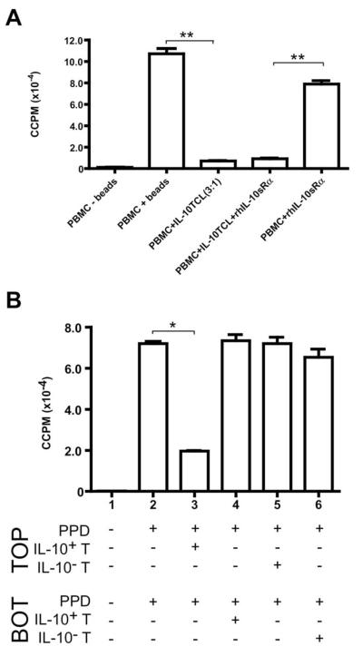
Fig. 4.
Cell contact, but neither IL-10 nor TGF-β, is required for IL-10+ T–cell–mediated suppression in vitro. (A) IL-10+ T cells were expanded with anti-CD3/CD28 beads, rhIL-2 and rhIL-15 for two rounds of stimulation; cells were used at day 9 of RII. Autologous naive PBMCs (1 × 10⁶ cells/condition) were activated in the presence (+) or absence (−) of anti-CD3/CD28 beads. IL-10+ T cells were titrated into wells with naive PBMCs at a 3:1 ratio (PBMCs: IL-10+ T cells). rhIL-10sRα (1 μg/mL) and/or rhTGF-βsRII (100 ng/mL) were added to cultures where indicated. At the peak response (day 3), 100-μl duplicates were harvested into 96-well plates and pulsed with 0.5 μCi 3[H]-thymidine. Results are expressed as ccpm; error bars represent SE. (B) IL-10+ and IL-10− T cells (selected and sorted from the same donor) were stimulated with PPD, IL-2 and IL-15 for 13 days. Autologous naive PBMCs (1 × 10⁶/ chamber) were added to the top and bottom chambers of transwells (separated by a porous membrane in 24-well plates), with or without PPD (50 μg/mL) in both chambers. Resting day 13 PPD-specific IL-10+ or IL-10− T cells were added to chambers where indicated at 3 × 10⁵ cells/chamber. On day 7, 100-μl duplicates were harvested into 96-well plates and pulsed with 0.5 μCi 3[H]-thymidine. Results are expressed as ccpm; error bars represent standard error. *p < 0.01; **p < 0.001. Data representative of three experiments (one distinct individual donor per experiment). TCL, T-cell line; Top, top well; bot, bottom well.

