Fig. 5.
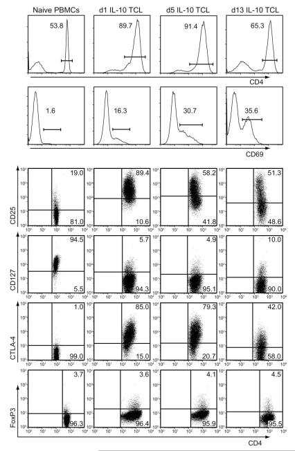
Anti-CD3/CD28 stimulated IL-10+ T cells have an activated cell phenotype. IL-10+ T cells (selected from human PBMCs) were expanded by stimulation with anti-CD3/CD28 (in the presence of IL-2 and IL-15) for one round, and then frozen. Thawed cells were subsequently washed and reactivated with anti-CD3/CD28 for 1, 5, or 13 days; stains depicted show autologous naive PBMC controls, d1 IL-10 TCL, day 5 IL-10 TCL, and day 13 IL-10 TCL. These cells were harvested and stained at each time point. Top two row histograms represent cell-surface staining for CD4 and CD69. Dot plots show CD4-gated cell surface stains for CD25 (third row), CD127 (fourth row), and CTLA-4 (fifth row). Bottom row shows dot plots for CD4-gated, intracellularly stained FoxP3 expression. Protein expression was measured by flow cytometry (BD FACS Scan) and data were analyzed with FlowJo software. Numbers represent the percentage expression of the described protein populations on the IL-10+ T-cell line (TCL) at indicated time points. Data are representative of two experiments.

