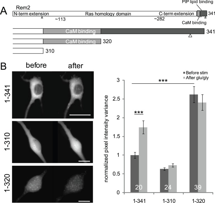
Figure 3. The Rem2 C-terminus directs redistribution.
(A) Schematic of the Rem2 protein. The last 30 residues of the C-terminal extension contain a previously identified PIP lipid binding domain as well as a calmodulin binding site. Small triangles in the N-and C-termini represent 14-3-3 binding sites. We created truncated Rem2 proteins ending at residues 310 and 320 to determine if these interaction sites are relevant for Rem2 redistribution. (B) Effect of Rem2 truncations on redistribution. (Left panel) Full-length Rem2 (top) redistributed into puncta on glutamate/glycine stimulation. 1–310 Rem2 (center) showed no redistribution upon stimulation, while 1–320 Rem2 (bottom) formed puncta constitutively. Scale bars indicate 5 µm. (Right panel) WT Rem2 shows a significant difference in pixel intensity variance after stimulation (normalized mean (± SEM) pixel value variance before stimulation, 1.00±0,08; after, 1.74±0.17; paired t-test p<0.001). Additionally, 1–320 Rem2 distribution is different from WT before but not after stimulation (one-way ANOVA followed by Bonferroni’s test; before, p<0.001; after, p = 0.19). Pixel intensity variances were normalized to the variance of full-length Rem2 before stimulation.

