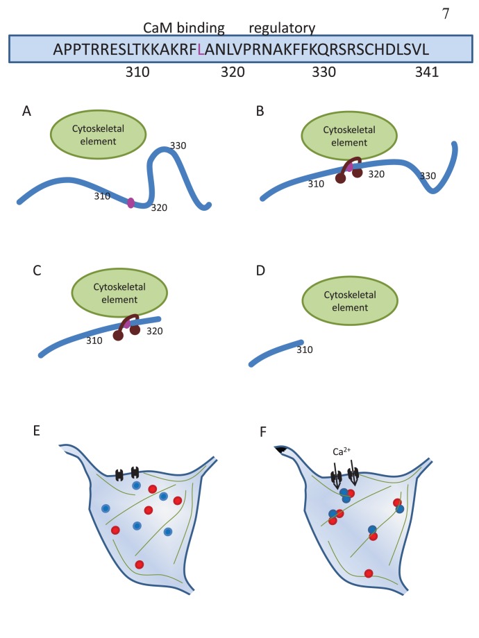
Figure 7. Possible mechanism underlying Rem2 activity-dependent redistribution.
Top: Representation of the C-terminus of Rem2, with the putative CaM-binding determinant leucine residue highlighted in pink. (A) In the absence of stimulation, Rem2 residues 320–330 (regulatory region) may allosterically inhibit Rem2 association with a putative cytoskeletal regulatory element, and consequently no Rem2 puncta are observed. (B) Upon stimulation, Ca2+-CaM may bind the region around residue L317, allowing redistribution to occur by association with the cytoskeleton. (C) The Rem2 mutant 1–320 lacks the regulatory region. This would result in constitutive association with CaM and constitutive puncta. (D) The Rem2 mutant 1–310 lacks the CaM-binding region, and would not redistribute to puncta unless CaMKII is overexpressed. (E) Under basal conditions, Rem2 (blue circles) and CaMKII (red circles) are diffusely distributed in neurons. (F) Upon neuronal stimulation, Ca2+ influx through the NMDAR (black brackets) leads to aggregation of Rem2 and CaMKII, possibly at cytoskeletal elements (green lines).

