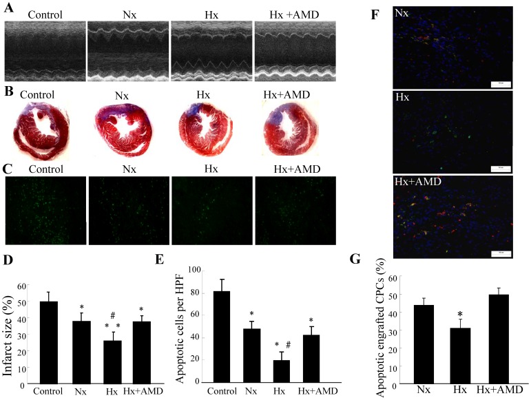
Figure 4. Effect of hypoxia preconditioned CPCs in treating MI.
CPCs were cultured under either normoxia (Nx) or hypoxia (Hx) for six hours with or without AMD3100 (CXCR4-selective antagonist, 5 µg/mL) and then intramyocardially injected into mice after surgical MI. Mice in the control group were administered DMEM. (A) Echocardiographic measurements were performed seven days after CPC transplantation. Representative M-mode images at the level of papillary muscles were recorded. (B) Representative Masson’s trichrome staining of transverse heart sections seven days after coronary ligation and CPC administration. (C) Representative phase micrographs show TUNEL-positive apoptotic cells in bordering myocardium adjacent to the infarct zone seven days after coronary ligation (×200 magnification). (D) Quantitative analysis of infarct size. Values are expressed as mean ± SD. n = 5. *P<0.05 vs. control group, **P<0.01 vs. control group, # P<0.05 vs. normoxia (Nx) & hypoxia (Hx) + AMD group. (E) Quantitative analysis of apoptotic cells in bordering myocardium. Values are expressed as mean ± SD. n = 5. *P<0.01 vs. control group, # P<0.01 vs. normoxia (Nx) & hypoxia (Hx) + AMD group. (F) Representative TUNEL+ apoptotic engrafted CPCs in heart tissues which were collected 72 h after cell transplantation (×200 magnification). (G) The percentage of apoptotic engrafted CPCs. The number of apoptotic engrafted CPCs was assessed by double positive of GFP+/TUNEL+ (yellow). Values are expressed as mean ± SD. n = 5. *P<0.01 vs. normoxia (Nx) group & hypoxia (Hx) + AMD group.

