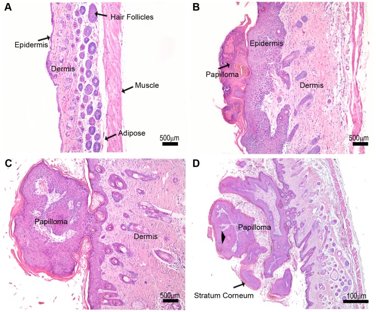
Figure 1. Tumor morphology changes with tumor progression.
(A) Control –The characteristic organization and thickness of epidermis and dermis are observed in a section of normal skin from a control mouse. (B) Phase I –This phase is characterized by epidermal proliferation, a pronounced thickening of the dermis and the formation of a papilloma. (C) Phase II - The dermis is thicker and the papilloma is more prominent. (D) Phase III - Keratin cysts (*) are observed, as well as a thickening of the stratum corneum.

