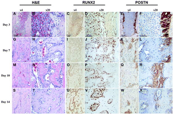
Figure 2.

Histologic evaluation of alveolar socket healing sites over time. Hematoxylin and eosin (H&E), RUNX2, and POSTN immunohistochemistry for tooth extraction site healing at 3, 7, 10, and 14 days (left panels, original magnification ×4; right panels, original magnification ×20). A and B) A clearly visible blood clot is noticeable at day 3. C and D) A significant number of RUNX2-positive cells are noticed within the alveolar socket populating the clot. Eand F) Remnants of the periodontal ligament can be clearly depicted by its strong POSTN staining at day 3. G through L) At day 7, the cell density in the defect area is higher and the POSTN- and RUNX2-positive cells start colocalizing within these areas. M through R) At day 10, the defect site seems to be filled by a condensed mesenchymal tissue. S through X) Finally, by day 14, an integration of the newly formed bone to the original socket walls is noticed.
