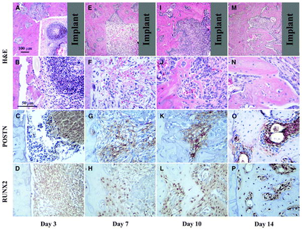
Figure 3.

Healing response for peri-implant repair sites at 3, 7, 10, and 14 days. A through D) Initially, inflammatory cells seem to dominate the defect area as depicted at day 3. E through H) By day 7, a loose fibrous connective tissue fills the defect and clear POSTN and RUNX2 staining is present. I through L) At day 10, RUNX2-positive cells are abundant and POSTN is gradually limited to the more immature tissue areas. M through P) Similar to the tooth extraction healing sites, at day 14, an integration of the newly formed bone to the walls of the defect is clear. Gray color in the top panels represents the implant location area (top panels, original magnification ×4; second, third, and fourth panels and rows, original magnification ×20).
