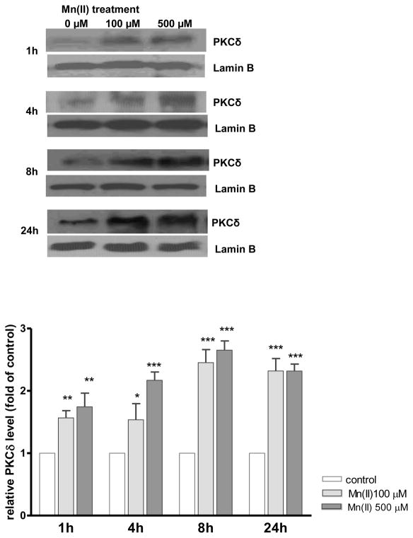
Fig. 6. Mn(II)-induced astrocytic PKCδ nuclear translocation.
Cells were treated with 100 μM or 500 μM of Mn(II) for 1, 4, 8 or 24 h. Next, the nuclear-rich fraction was isolated with the Nuclear/cytosol fractionation kit following the manufacturer’s protocol (BioVison, Mountain View, CA, USA) and analyzed for PKCδ and the nuclear marker Lamin B by western blotting. Data represent the mean ± S.D. from 3 independent sets of cultures, each performed in triplicate; *p < 0.05, **p < 0.01, ***p < 0.001 control vs. Mn(II)-exposed cells (one-way ANOVA followed by post hoc Tukey’s test).

