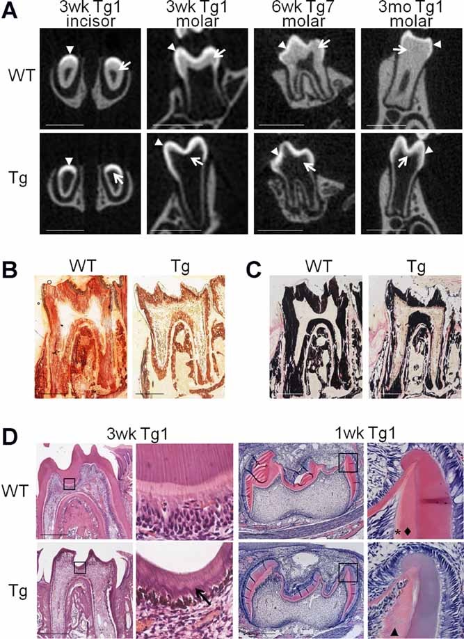
Fig. 2.

Defective dentin formation in Col1a1-Trps1 mice. (A) µCT images (cross sections) of WT and transgenic teeth (Tg1 and Tg7 line) demonstrating a diminished dentin layer in transgenic mice. Arrowheads: enamel, arrows: dentin. Transgenic animals were fed with soft food to assure proper nutrition. Scale bars = 1 mm. (B) Alizarin red staining of undecalcified molar sections (3 weeks old) demonstrating reduced dentin layer in transgenic teeth. Scale bars = 500 µm. (C) Von Kossa staining of undecalcified molar sections (3 weeks old) demonstrating reduced dentin layer in transgenic teeth. Scale bars = 500 µm. (D) Histological analyses of molars showing abnormal dentin formation in transgenic mice. In the dentin of 3-week-old (3wk) transgenic animals (erupted teeth) an irregular mineralization front is observed (arrow). In the 1-week-old (1wk) WT animals (unerupted teeth) two distinctive layers of predentin and dentin are present (asterisk and diamond, respectively), while in transgenic animals only one layer is present (arrowhead). Black boxes indicate areas magnified on the images to the right. Scale bars = 500 µm.
